Smooth muscles. Smooth muscles Smooth muscle cells
Muscle tissues are tissues that differ in structure and origin, but have a common ability to contract. They consist of myocytes - cells that can perceive nerve impulses and respond to them with a contraction.
Properties and types of muscle tissue
Morphological features:
- Elongated form of myocytes;
- longitudinally placed myofibrils and myofilaments;
- mitochondria are located near contractile elements;
- polysaccharides, lipids and myoglobin are present.
Properties of muscle tissue:
- contractility;
- excitability;
- conductivity;
- extensibility;
- elasticity.
There are the following types muscle tissue depending on morphofunctional features:
- Striated: skeletal, cardiac.
- Smooth.
Histogenetic classification divides muscle tissue into five types depending on the embryonic source:
- Mesenchymal - desmal germ;
- epidermal - skin ectoderm;
- neural - neural plate;
- coelomic - splanchnotomes;
- somatic - myotome.
Of 1-3 species, smooth muscle tissues develop, 4, 5 give striated muscles.
The structure and function of smooth muscle tissue
Consists of individual small spindle-shaped cells. These cells have a single nucleus and thin myofibrils that extend from one end of the cell to the other. Smooth muscle cells are combined into bundles, consisting of 10-12 cells. This association arises due to the peculiarities of smooth muscle innervation and facilitates the passage of a nerve impulse to the entire group of smooth muscle cells. Smooth muscle tissue contracts rhythmically, slowly and for a long time, while being able to develop great strength without significant energy expenditure and without fatigue.
In lower multicellular animals, all muscles are composed of smooth muscle tissue, while in vertebrates it is part of the internal organs (except the heart).
The contractions of these muscles do not depend on the will of the person, that is, they occur involuntarily.
Functions of smooth muscle tissue:
- Maintaining stable pressure in hollow organs;
- regulation of blood pressure;
- peristalsis of the digestive tract, movement of contents along it;
- bladder emptying.
The structure and function of skeletal muscle tissue

Consists of long and thick fibers 10-12 cm long. Skeletal muscles are characterized by voluntary contraction (in response to impulses coming from the cerebral cortex). The speed of its contraction is 10-25 times higher than in smooth muscle tissue.
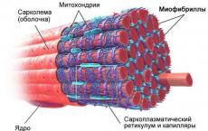
The muscle fiber of the striated tissue is covered with a sheath - the sarcolemma. Under the membrane is the cytoplasm with a large number of nuclei located along the periphery of the cytoplasm, and contractile filaments - myofibrils. The myofibril consists of successively alternating dark and light areas (discs) with different light refractive index. Using an electron microscope, it was found that the myofibril consists of protofibrils. Thin protofibrils are built from a protein - actin, and thicker ones - from myosin.
When the fibers contract, excitation of contractile proteins occurs, thin protofibrils glide over thick ones. Actin reacts with myosin to form a single actomyosin system.
Functions of skeletal muscle tissue:
- Dynamic - movement in space;
- static - maintaining a certain position of body parts;
- receptor - proprioceptors that perceive irritation;
- depositing - liquid, minerals, oxygen, nutrients;
- thermoregulation - relaxation of muscles with an increase in temperature to expand blood vessels;
- facial expressions - to convey emotions.
The structure and function of cardiac muscle tissue
 cardiac muscle tissue
 The myocardium is built from cardiac muscle and connective tissue, with vessels and nerves. Muscle tissue refers to striated muscles, the striation of which is also due to the presence of different types of myofilaments. The myocardium is made up of fibers that are interconnected and form a mesh. These fibers include single or binuclear cells that are arranged in a chain. They are called contractile cardiomyocytes.
Contractile cardiomyocytes are 50 to 120 micrometers long and up to 20 microns wide. The nucleus here is located in the center of the cytoplasm, in contrast to the nuclei of striated fibers. Cardiomyocytes have more sarcoplasm and fewer myofibrils than skeletal muscle. There are many mitochondria in the cells of the heart muscle, since continuous heartbeats require a lot of energy.
The second type of myocardial cells are conductive cardiomyocytes, which form the conduction system of the heart. Conductive myocytes provide impulse transmission to contractile muscle cells.
Functions of cardiac muscle tissue:
- Pump house;
- provides blood flow in the bloodstream.
Components of the contractile system
The structural features of muscle tissue are determined by the functions performed, the ability to receive and conduct impulses, and the ability to contract. The contraction mechanism consists in the coordinated work of a number of elements: myofibrils, contractile proteins, mitochondria, myoglobin.
In the cytoplasm of muscle cells there are special contractile filaments - myofibrils, the contraction of which is possible with the friendly work of proteins - actin and myosin, as well as with the participation of Ca ions. Mitochondria supply all processes with energy. Also, energy reserves form glycogen and lipids. Myoglobin is necessary for the binding of O 2 and the formation of its reserve for the period of muscle contraction, since during contraction there is compression of the blood vessels and the supply of O 2 to the muscles is sharply reduced.
Table. Correspondence between the characteristics of muscle tissue and its type
| Type of fabric | Characteristic |
|---|---|
| smooth muscle | Included in the walls of blood vessels |
| Structural unit - smooth myocyte | |
| Decreases slowly, unconsciously | |
| There is no transverse striation | |
| Skeletal | Structural unit - multinuclear muscle fiber |
| Characterized by transverse striation | |
| Decreases quickly, consciously |
Where is muscle tissue located?
Smooth muscles are an integral part of the walls of internal organs: the gastrointestinal tract, the genitourinary system, and blood vessels. They are part of the capsule of the spleen, skin, sphincter of the pupil.
Skeletal muscles occupy about 40% of the human body weight, they are attached to the bones with the help of tendons. This tissue consists of skeletal muscles, muscles of the mouth, tongue, pharynx, larynx, upper esophagus, diaphragm, mimic muscles. Also, striated muscle is located in the myocardium.
How is skeletal muscle fiber different from smooth muscle tissue?
The fibers of striated muscles are much longer (up to 12 cm) than the cellular elements of smooth muscle tissue (0.05-0.4 mm). Also, skeletal fibers have transverse striation due to the special arrangement of actin and myosin filaments. This is not the case for smooth muscles.
There are many nuclei in muscle fibers, and the contraction of the fibers is strong, fast and conscious. Unlike smooth muscles, smooth muscle tissue cells are mononuclear, able to contract at a slow pace and unconsciously.
Ministry of Education and Science of the Russian Federation
Federal State Budgetary Educational Institution of Higher Professional Education
«***»
ABSTRACT
at the rate
"basics of anatomy and physiology"
on the topic
"Smooth muscles. Structure, functions, reduction mechanism "
Lead teacher:
Art. teacher **
Work completed:
Group student **
Evaluation based on the results of the defense of the abstract:
_____________________
"___" __________20__
Moscow 2013
- Introduction……………………………………………………………………………….2
- The structure of smooth muscles……………………………………………………………...3
- Smooth muscle functions ………………………………………………………………...5
- Reduction mechanism………………………………………………………………..8
- Excitatory and inhibitory mediators secreted in the neuromuscular junctions of smooth muscles…………………………………………………………...10
- Conclusion…………………………………………………………………………...11
- List of used literature………………………………………………….12
Introduction
Muscles or Muscles (from lat. musculus muscle) organs bodies of animals and humans, consisting of an elastic, elastic muscle tissue , capable of contracting under the influencenerve impulses. Designed to perform various actions: body movements, contractions of voice ligaments, breathing . Muscles allow you to move parts of the body and express thoughts and feelings in actions. A person performs any movement from such simple things as blinking or smile , to thin and energetic, such as we see in jewelers or athletes due to the ability of muscle tissue to contract.
Smooth muscles are an integral part of some internal organs and are involved in providing the functions performed by these organs. In particular, they regulate the patency of the bronchi for air, blood flow in various organs and tissues, the movement of fluids and chyme (in the stomach, intestines, ureters, urinary and gall bladders), expel the fetus from the uterus, dilate or narrow the pupils (due to the reduction of radial or circular muscles of the iris), change the position of the hair and skin relief.
The structure of smooth muscles
There are three groups of smooth (non-striated) muscle tissues: mesenchymal, epidermal and neural.
Muscle tissue of mesenchymal origin.
Stem cells and progenitor cells in smooth muscle tissue at the stages of embryonic development have not yet been accurately identified. Apparently, they are related to the mechanocytes of the tissues of the internal environment. Probably, in the mesenchyme they migrate to the sites of organ anlage, being already determined. Differentiating, they synthesize components of the matrix and collagen of the basement membrane, as well as elastin. In definitive cells (myocytes), the synthetic ability is reduced, but does not disappear completely. Smooth myocyte fusiform cell 20 500 µm long, 5 8 µm wide. The nucleus is rod-shaped, located in its central part. When a myocyte contracts, its nucleus bends and even twists. Organelles of general importance, among which there are many mitochondria, are concentrated near the poles of the nucleus (in the endoplasm). The Golgi apparatus and the granular endoplasmic reticulum are poorly developed, which indicates a low activity of synthetic functions. Ribosomes are mostly located
free. Myocytes are combined into bundles, between which there are thin layers of connective tissue. Reticular and elastic fibers surrounding the myocytes are woven into these layers. In the layers are blood vessels and nerve fibers. The terminals of the latter do not end directly on the myocytes, but between them. Therefore, after the arrival of a nerve impulse, the mediator spreads diffusely, exciting many cells at once.
Smooth muscle tissue of mesenchymal origin is represented mainly in the walls of blood vessels and many tubular internal organs, and also forms separate small muscles (ciliary).
Muscular tissue of epidermal origin.Myoepithelial cells develop from the epidermal bud. They are found in the sweat, mammary, salivary, and lacrimal glands and share common precursors with
their secretory cells. Myoepithelial cells are directly adjacent to the epithelial cells proper and have a basement membrane in common with them. During regeneration, those and other cells are also restored from common undifferentiated precursors. Most myoepithelial cells are stellate in shape. These cells are often called basket cells: their processes cover the terminal sections and small ducts of the glands.
The nucleus and organelles of general importance are located in the cell body, and the contractile apparatus is located in the processes, organized, as in the cells of the mesenchymal type of muscle tissue.
Muscle tissue of neural origin.
The myocytes of this tissue develop from the cells of the neural rudiment as part of the inner wall of the eyecup. The bodies of these cells are located in the epithelium of the posterior surface of the iris. Each of them has a process that goes into the thickness of the iris and lies parallel to its surface. The process contains a contractile apparatus, organized in the same way as in all smooth myocytes. Depending on the direction of the processes (perpendicular or parallel to the edge of the pupil), myocytes form two muscles: constricting and dilating the pupil.
It should be remembered that the composition of smooth muscle tissue, regardless of its origin, also includes specific constituent elements that are directly related to the mechanism of contraction directly, these are myofibrils. The structure, which includes "contractile" proteins, which are called actin and myosin.
Myosin - protein of contractile muscle fibers. Its content in muscles is about 40% of the mass of all proteins (for example, in other tissues it is only 1-2%). The myosin molecule is a long filamentous rod, as if two ropes were woven together, forming two pear-shaped heads at one end.
Actin so is the protein of contractile muscle fibers, much smaller in size than myosin, and occupying only 15-20% of the total mass of all proteins. It consists of two threads woven into a rod, with grooves.
Smooth muscle functions
Smooth muscles, like skeletal muscles, are excitable, conductive, and contractile. Unlike skeletal muscles, which have elasticity, smooth are plastic (capable of maintaining the length given to them by stretching for a long time without increasing tension). This property is important for the function of depositing food in the stomach or fluids in the gallbladder or bladder.
Features of excitability smooth muscle fibers are to some extent associated with their low transmembrane potential (E 0 = 30-70 mV). Many of these fibers are automatic. The duration of the action potential in them can reach tens of milliseconds. This happens because the action potential in these fibers develops mainly due to the entry of calcium into the sarcoplasm from the intercellular fluid through the so-called slow Ca 2+ channels.
Visceral smooth muscles are characterized by unstable membrane potential. Fluctuations in membrane potential, regardless of nerve influences, cause irregular contractions that maintain the muscle in a state of constant partial contraction - tone. The tone of smooth muscles is clearly expressed in the sphincters of hollow organs: the gallbladder, bladder, at the junction of the stomach into the duodenum and the small intestine into the colon, as well as in the smooth muscles of small arteries and arterioles. The membrane potential of smooth muscle cells is not a reflection of the true value of the resting potential. With a decrease in membrane potential, the muscle contracts, with an increase in , it relaxes. During periods of relative rest, the value of the membrane potential is on average 50 mV. In visceral smooth muscle cells, slow wave-like fluctuations of the membrane potential of several millivolts are observed, as well as an action potential (AP). The value of PD can vary over a wide range. In smooth muscles, the duration of AP 50 250 ms; There are PDs of various shapes. In some smooth muscles, such as the ureter, stomach, and lymphatics, APs have a prolonged plateau during depolarization, reminiscent of the potential plateau in myocardial cells. Plate-like APs ensure the entry into the cytoplasm of myocytes of a significant
the amount of extracellular calcium, which subsequently participates in the activation of contractile proteins of smooth muscle cells. The ionic nature of smooth muscle AP is determined by the features of the channels of the smooth muscle cell membrane. Ca2+ ions play the main role in the mechanism of AP occurrence. Calcium channels of the membrane of smooth muscle cells pass not only Ca2+ ions, but also other doubly charged ions (Ba 2+, Mg2+), as well as Na+. The entry of Ca2+ into the cell during PD is necessary to maintain tone and develop contraction; therefore, blocking the calcium channels of the smooth muscle membrane, which leads to a restriction of Ca2+ ion entry into the cytoplasm of myocytes of internal organs and vessels, is widely used in practical medicine to correct the motility of the digestive tract and vascular tone in the treatment of patients with hypertension.
Speed excitationin smooth muscle cells small 2-10 cm/s. Unlike skeletal muscles, excitation in a smooth muscle can be transmitted from one fiber to another nearby. This conduction occurs due to the presence of nexuses between smooth muscle fibers (areas of contact between twocell membranes, where are the channels for the exchange ions and micromolecules) , which have low resistance to electric current and ensure the exchange between Ca cells 2+ and other molecules. As a result, smooth muscle has the properties of a functional syncytium (represents several cells that have merged with each other and contain several nuclei).
Contractility smooth muscle fibers is characterized by a long latent period (the time between the onset of the stimulus and the occurrence of a response) (0.25-1.00 s) and a long duration (up to 1 min) of a single contraction. Smooth muscles have a low contraction force, but are able to stay in tonic contraction for a long time without developing fatigue. This is due to the fact that smooth muscle consumes 100-500 times less energy than skeletal muscle to maintain tonic contraction (prolonged contraction). Therefore, the ATP reserves consumed by the smooth muscle have time to recover even during contraction, and the smooth muscles of some body structures are in a state of tonic contraction all their lives (are actually a kind of tetanic contractions,
representing a long-term shortening of the muscles and causing mainly muscle tone - a constant slight muscle tension that occurs in muscle tissue at rest. This constant tension of muscle tissue takes place even in the state of sleep).
Relationship of excitation with contraction. It is more difficult to study the relationship between electrical and mechanical manifestations in visceral smooth muscle than in skeletal or cardiac muscle, since visceral smooth muscle is in a state of continuous activity. Under conditions of relative rest, a single AP can be registered. The contraction of both skeletal and smooth muscles is based on the sliding of actin in relation to myosin, where the Ca2+ ion performs a trigger function (the ability to stay in one state for a long time).
A unique feature of visceral smooth muscle is its response to stretch. Smooth muscle contracts in response to stretch. This is due to the fact that stretching reduces the membrane potential of cells, increases the frequency of AP and, ultimately, smooth muscle tone. In the human body, this property of smooth muscles is one of the ways to regulate the motor activity of internal organs. For example, when the stomach is full, it expands. walls . An increase in the tone of the stomach wall in response to its stretching contributes to the preservation of the volume of the organ and better contact of its walls with the incoming food. In blood vessels, the stretch created by fluctuations in blood pressure is the main factor in myogenic self-regulation of vascular tone. Finally, stretching of the muscles of the uterus by a growing fetus is one of the reasons for the onset of labor.
Reduction mechanism
Conditions for smooth muscle contraction.
The most important feature of smooth muscle fibers is that they are excited under the influence of numerous stimuli. Skeletal muscle contraction is normally initiated only by a nerve impulse passing to the neuromuscular synapse. Smooth muscle contraction can be caused both by a nerve impulse and by biologically active substances (hormones, many neurotransmitters, some metabolites), as well as by physical factors, such as stretching. In addition, smooth muscle contraction can occur spontaneously due to automation.
The very high reactivity of smooth muscles, their ability to respond to contractions to the action of various factors create significant difficulties for the correction of violations of the tone of these muscles in medical practice. This can be seen in the example of bronchial asthma, arterial hypertension and other diseases that require correction of the contractile activity of smooth muscles.
In the molecular mechanism of smooth muscle contraction, there are also a number of differences from skeletal muscle contraction. Actin and myosin filaments in smooth muscle fibers are less ordered than in skeletal ones, and therefore smooth muscle does not have transverse striation. There is no troponin protein in actin filaments of smooth muscle, and actin molecular centers are always open for interaction with myosin heads. For this interaction to occur, splitting of the ATP molecule and transfer of phosphate to the myosin heads is necessary. This is followed by the rotation of the myosin heads, in which the actin filaments are drawn in between the myosin filaments and contraction occurs.
Phosphoration of myosin heads occurs with the help of the enzyme myosin light chain kinase, and dephosphorylation occurs with the help of myosin light chain phosphatase. If the activity of myosin phosphatase predominates over the kinase, then the myosin heads are dephosphorylated, the actin-myosin bond is broken, and the muscles relax.
Therefore, for smooth muscle contraction to occur, an increase in the activity of myosin light chain kinase is required. Its activity is regulated by the level of Ca 2+ in the sarcoplasm. When a smooth muscle fiber is stimulated, the calcium content in its sarcoplasm increases. This increase is due to the intake of Ca 2+ from two sources: 1) intercellular space; 2) sarcoplasmic reticulum. Further, calcium ions form a complex with the calmodulin protein, which activates myosin kinase.
Sequence of processes leading to the development of smooth muscle contraction: Ca input 2+ into the sarcoplasm Activation of calmodulin Activation of myosin light chain kinase Phosphorylation of myosin heads Binding of myosin heads to actin and turning of the heads, in which the actin filaments are pulled between the myosin filaments.
Conditions necessary for smooth muscle relaxation.
- Decrease (up to 10 -7 M/l and less) Ca content 2+ in sarcoplasm;
- Decomposition of the 4 Ca complex 2+ - calmodulin, leading to a decrease in the activity of myosin light chain kinase, dephosphorylation of myosin heads, leading to rupture of bonds between actin and myosin filaments
After that, the elastic forces cause a relatively slow recovery of the original length of the smooth muscle fiber, its relaxation.
Excitatory and inhibitory mediators secreted at the neuromuscular junctions of smooth muscles.
The most important mediators secreted by the autonomic nerves that innervate smooth muscles are acetylcholine and norepinephrine, but they are never released by the same nerve fibers. Acetylcholine acts as an excitatory mediator for the smooth muscles of some organs, and acts as an inhibitory agent for the smooth muscles of other organs. If acetylcholine excites the muscle fiber, norepinephrine usually inhibits it. Conversely, if acetylcholine inhibits the fiber, norepinephrine tends to excite it. But why are there such different reactions? The answer is that acetylcholine and norepinephrine either excite or inhibit smooth muscle by first binding to a receptor protein on the surface of the muscle cell membrane. Some of these receptor proteins are excitatory receptors while others are inhibitory receptors. Therefore, the type of receptor determines how the smooth muscle will react with inhibition or excitation, as well as which of the two mediators (acetylcholine or norepinephrine) will exhibit an excitatory or inhibitory effect.
Conclusion
Many smooth muscles in the skin, they are located at the base of the hair bag. By contracting, these muscles raise the hair and squeeze out fat from the sebaceous gland. In the eye around the pupil are smooth circular and radial muscles. They work all the time: in bright light, the circular muscles constrict the pupil, and in the dark, the radial muscles contract and the pupil expands. In the walls of all tubular organs - the respiratory tract, blood vessels, digestive tract, urethra, etc. - there is a layer of smooth muscles. Under the influence of nerve impulses, it is reduced. Due to the contraction and relaxation of the smooth cells of the walls of blood vessels, their lumen either narrows or expands, which contributes to the distribution of blood in the body. The smooth muscles of the esophagus, contracting, push a lump of food or a sip of water into the stomach. Complex plexuses of smooth muscle cells are formed in organs with a wide cavity - in the stomach, bladder, uterus. The contraction of these cells causes compression and narrowing of the lumen of the organ. The strength of each cell contraction is negligible, because. they are very small. However, the addition of the forces of entire beams can create a contraction of enormous force. Powerful contractions create a sensation of intense pain. Excitation in smooth muscles spreads relatively slowly, which leads to slow long-term contraction of the muscle and an equally long period of relaxation. Muscles are also capable of spontaneous rhythmic contractions. The stretching of the smooth muscles of the hollow organ when filled with its contents immediately leads to its contraction - this ensures that the contents are pushed further.
This list of examples of smooth muscle in the human body can be continued indefinitely, thus showing the enormous importance of smooth muscle.
List of used literature
- Histology. Yu.I. Afanasiev, N.A. Yurina, E.F. Kotovsky, 2002
- Atlas of histology and embryology.I.V. Almazov, L.S. Sutulov, 1978
- Human anatomy. M.F. Ivanitsky, 2008
- Anatomy. I.V. Gaivoropsky, G.I. Nichiporuk, 2006
- Human physiology. A.A. Semenovich, 2009
PAGE \* MERGEFORMAT 1
Smooth muscles in the body of higher animals and humans are located in the internal organs, in the vessels and in the skin. Smooth musclesare able to carry out relatively slow movements and long tonic contractions.
Relatively slow, often rhythmic contractions smooth muscles the walls of hollow organs: the stomach, intestines, ducts of the digestive glands, bladder, gallbladder, etc. - provide movement and ejection of the contents of these hollow organs. An example is the pendulum and peristaltic movements of the intestinal musculature.
Prolonged tonic contractions of smooth muscles are especially pronounced in the sphincters of hollow organs; their tonic contraction prevents the release of the contents of the organ. This ensures the accumulation of bile in the gallbladder and urine in the bladder, the formation of feces in the rectum, etc.
The smooth muscles of the walls of blood vessels, especially arteries and arterioles, also have a pronounced tone. The tone of the muscle layer of the walls of the artery regulates the size of their lumen, and thus the level of blood pressure and blood supply to the organs.
The tone and motor function of smooth muscles are regulated by impulses coming through the autonomic nerves and by humoral influences.
Main functions smooth muscles:
- in hollow organs (ureter, intestines, etc.) they maintain pressure;
- slow contraction of smooth muscles causes undulating peristalsis of hollow organs,
- which ensures the promotion of their contents and the emptying of organs;
- change the lumen of blood vessels, thereby regulating the pressure in them;
- smooth muscles located in the skin at the base of the hair bags, when contracted, raise the hair and squeeze out fat from the sebaceous glands;
- in the eyes, smooth muscles provide constriction and expansion of the pupil, determine the thickness of the lens.
feature smooth muscle is:
- slow contraction and relaxation (tens of seconds);
- involuntary nature of the contraction (regardless of the will of the person).
Smooth muscle properties
Smooth muscle plasticity
Important properties of smooth muscle is its great plasticity, i.e., the ability to maintain the length given by stretching without changing the stress. The difference between skeletal muscle, which has little plasticity, and smooth muscle, with well-defined plasticity, is easily detected if they are first slowly stretched, and then the tensile load is removed. Skeletal muscle immediately shortened after the load is removed. In contrast, the smooth muscle after the removal of the load remains stretched until, under the influence of some kind of irritation, its active contraction occurs.
The property of plasticity is very important for the normal activity of the smooth muscles of the walls of hollow organs, such as the bladder: due to the plasticity of the smooth muscles of the walls of the bladder, the pressure inside it changes relatively little with different degrees of filling.

Excitability and arousal
Smooth muscles less excitable than skeletal ones: their thresholds of irritation are higher, and the chronaxy is longer. The action potentials of most smooth muscle fibers have a small amplitude (about 60 mV instead of 120 mV in skeletal muscle fibers) and a long duration - up to 1-3 seconds. On rice. 151 shows the action potential of a single fiber of the uterine muscle.
The refractory period lasts for the entire period of the action potential, i.e. 1-3 seconds. The rate of excitation conduction varies in different fibers from a few millimeters to several centimeters per second.
There are a large number of different types of smooth muscle in the body of animals and humans. Most of the hollow organs of the body are lined with smooth muscles that have a sensitial type of structure. The individual fibers of such muscles are very closely adjacent to each other and it seems that morphologically they form a single whole.
Smooth muscle irritants . One of the important physiologically adequate stimuli of smooth muscles is their rapid and strong stretching. The latter causes depolarization of the muscle fiber membrane and the occurrence of a propagating action potential.
Smooth muscles are found in hollow organs, blood vessels, and skin. Smooth muscle fibers do not have transverse striation. The cells shorten as a result of the relative sliding of the filaments. The sliding speed and the rate of splitting of adenosine triphosphate are 100-1000 times less than in. Due to this, smooth muscles are well adapted for long-term stable contraction without fatigue, with less energy expenditure.
Smooth muscles are an integral part of the walls of a number of hollow internal organs and are involved in providing the functions performed by these organs. In particular, they regulate blood flow in various organs and tissues, bronchial patency for air, movement of fluids and chyme (in the stomach, intestines, ureters, urinary and gallbladder), contraction of the uterus during childbirth, pupil size, skin relief.
Smooth muscle cells are spindle-shaped, 50-400 microns long, 2-10 microns thick (Fig. 5.6).
Smooth muscles are involuntary muscles, i.e. their reduction does not depend on the will of the macroorganism. Features of the motor activity of the stomach, intestines, blood vessels and skin to a certain extent determine the physiological characteristics of the smooth muscles of these organs.
Characteristics of smooth muscle
- It has automatism (the influence of the intramural nervous system is corrective)
- Plasticity - the ability to maintain length for a long time without changing tone
- Functional syncytium - individual fibers are separated, but there are special areas of contact - nexuses
- The value of the resting potential is 30-50 mV, the amplitude of the action potential is less than that of skeletal muscle cells
- Minimum "critical zone" (excitation occurs if a certain minimum number of muscle elements are excited)
- For the interaction of actin and myosin, the Ca 2+ ion is required, which comes from outside
- The duration of a single contraction is long
feature of smooth muscle- their ability to show slow rhythmic and long tonic contractions. Slow rhythmic contractions of the smooth muscles of the stomach, intestines, ureters and other hollow organs contribute to the movement of their contents. Prolonged tonic contractions of the smooth muscles of the sphincters of the hollow organs prevent the arbitrary release of their contents. The smooth muscles of the walls of blood vessels are also in a state of constant tonic contraction and affect the level of blood pressure and blood supply to the body.
An important property of smooth muscles is their mysticism, those. the ability to retain the shape caused by stretching or deformation. High plasticity of smooth muscles is of great importance for the normal functioning of organs. For example, the plasticity of the bladder makes it possible, when it is filled with urine, to prevent an increase in pressure in it without disturbing the process of urination.
Excessive stretching of smooth muscles causes them to contract. This occurs as a result of depolarization of cell membranes caused by their stretching, i.e. smooth muscles have automatism.
The contraction caused by stretch plays an important role in the autoregulation of blood vessel tone, the movement of the contents of the gastrointestinal tract, and other processes.
Rice. 1. A. Skeletal muscle fiber, cardiac muscle cell, smooth muscle cell. B. Skeletal muscle sarcomere. B. The structure of smooth muscle. D. Mechanogram of skeletal muscle and heart muscle.
Automatism in smooth muscles is due to the presence in them of special pacemaker (rhythm-setting) cells. In their structure, they are identical to other smooth muscle cells, but have special electrophysiological properties. Pacemaker potentials arise in these cells, depolarizing the membrane to a critical level.
Excitation of smooth muscle cells causes an increase in the entry of calcium ions into the cell and the release of these ions from the sarcoplasmic reticulum. As a result of an increase in the concentration of calcium ions in the sarcoplasm, contractile structures are activated, but the mechanism of their activation in a smooth fiber differs from the mechanism of activation in striated muscles. In a smooth cell, calcium interacts with the protein calmodulin, which activates myosin light chains. They connect to the active centers of actin in protofibrils and make a "stroke". Smooth muscles relax passively.
Smooth muscles are involuntary, and they do not depend on the will of the animal.
Physiological properties and features of smooth muscles
Smooth muscles, like skeletal muscles, have excitability, conductivity, and contractility. Unlike skeletal muscles, which have elasticity, smooth muscles have plasticity - the ability to maintain the length given to them during stretching for a long time without increasing tension. This property is important for the function of depositing food in the stomach or fluids in the gallbladder and bladder.
Features of the excitability of smooth muscle cells are to a certain extent associated with a low potential difference across the membrane at rest (E 0 = (-30) - (-70) mV). Smooth myocytes can be automatic and spontaneously generate an action potential. Such cells - pacemakers of smooth muscle contraction are found in the walls of the intestine, venous and lymphatic vessels.

Rice. 2. The structure of a smooth muscle cell (A. Guyton, J. Hall, 2006)
The duration of AP in smooth myocytes can reach tens of milliseconds, since AP develops in them mainly due to the entry of Ca 2+ ions into the sarcoplasm from the intercellular fluid through slow calcium channels.
The speed of AP conduction along the membrane of smooth myocytes is low - 2-10 cm/s. Unlike skeletal muscles, excitation can be transmitted from one smooth myocyte to others nearby. Such a transfer occurs due to the presence of nexuses between smooth muscle cells, which have low resistance to electric current and ensure the exchange of Ca 2+ ions and other molecules between cells. As a result, smooth muscle exhibits the properties of functional syncytium.
The contractility of smooth muscle cells is characterized by a long latent period (0.25-1.00 s) and a long duration (up to 1 min) of a single contraction. Smooth muscles develop a small force of contraction, but are able to stay in a tonic contraction for a long time without developing fatigue. This is due to the fact that smooth muscle consumes 100-500 times less energy than skeletal muscle to maintain tonic contraction. Therefore, the ATP reserves consumed by the smooth muscle have time to recover even during contraction, and the smooth muscles of some body structures are almost constantly in a state of tonic contraction. The absolute strength of smooth muscle is about 1 kg/cm 2 .
Mechanism of smooth muscle contraction
The most important feature of smooth muscle cells is that they are excited under the influence of numerous stimuli. under natural conditions, it is initiated only by a nerve impulse coming to. Contraction of a smooth muscle can be caused both by the influence of nerve impulses and by the action of hormones, neurotransmitters, prostaglandins, some metabolites, as well as by the influence of physical factors, such as stretching. In addition, excitation and contraction of smooth myocytes can occur spontaneously - due to automation.
The ability of smooth muscles to respond by contraction to the action of various factors will create significant difficulties for correcting violations of the tone of these muscles in medical practice. This can be seen in the examples of difficulties in the treatment of bronchial asthma, arterial hypertension, spastic colitis and other diseases that require correction of the contractile activity of smooth muscles.
The molecular mechanism of smooth muscle contraction also has a number of differences from the mechanism of skeletal muscle contraction. The actin and myosin filaments in smooth muscle cells are less ordered than in skeletal cells, and therefore smooth muscle does not have transverse striation. There is no troponin protein in actin filaments of smooth muscle, and actin centers are always open for interaction with myosin heads. At the same time, myosin heads are not energized at rest. In order for actin and myosin to interact, it is necessary to phosphorylate the myosin heads and give them an excess of energy. The interaction of actin and myosin is accompanied by the rotation of the myosin heads, in which the actin filaments are pulled between the myosin filaments and the contraction of the smooth myocyte occurs.
Phosphorylation of myosin heads is carried out with the participation of the enzyme myosin light chain kinase, and dephosphorylation is carried out with the help of phosphatase. If the activity of myosin phosphatase predominates over the activity of the kinase, then the myosin heads are dephosphorylated, the connection between myosin and actin is broken, and the muscle relaxes.
Therefore, in order for smooth myocyte contraction to occur, it is necessary to increase the activity of myosin light chain kinase. Its activity is regulated by the level of Ca 2+ ions in the sarcoplasm. Neurotransmitters (acetylcholine, noradrsnaline) or hormones (vasopressin, oxytocin, adrenaline) stimulate their specific receptor, causing dissociation of the G-protein, the a-subunit of which further activates the phospholipase C enzyme. cell membranes. IPG diffuses to the endoplasmic reticulum and, after interacting with its receptors, causes the opening of calcium channels and the release of Ca 2+ ions from the depot into the cytoplasm. An increase in the content of Ca 2+ ions in the cytoplasm is a key event for the initiation of smooth myocyte contraction. An increase in the content of Ca 2+ ions in the sarcoplasm is also achieved due to its entry into the myocyte from the extracellular medium (Fig. 3).
Ca 2+ ions form a complex with the calmodulin protein, and the Ca 2+ -calmodulin complex increases the kinase activity of myosin light chains.
The sequence of processes leading to the development of smooth muscle contraction can be described as follows: entry of Ca 2+ ions into the sarcoplasm - activation of calmodulin (by formation of the 4Ca 2 -calmodulin complex) - activation of myosin light chain kinase - phosphorylation of myosin heads - binding of myosin heads to actin and the rotation of the heads, in which the actin filaments are drawn in between the myosin filaments - contraction.

Rice. Fig. 3. Ways of entry of Ca 2+ ions into the sarcoplasm of a smooth muscle cell (a) and their removal from the sarcoplasm (b)
Conditions necessary for smooth muscle relaxation:
- a decrease (up to 10-7 M/l or less) in the content of Ca 2+ ions in the sarcoplasm;
- disintegration of the 4Ca 2+ -calmodulin complex, leading to a decrease in the activity of myosin light chain kinase - dephosphorylation of myosin heads under the influence of phosphatase, leading to a break in the bonds of actin and myosin filaments.
Under these conditions, elastic forces cause a relatively slow recovery of the original length of the smooth muscle fiber and its relaxation.
Smooth muscles in invertebrates and vertebrates
Smooth muscle contractions
Unlike striated muscles, smooth muscles are characterized by slow contraction, the ability to stay in a state of contraction for a long time, expending relatively little energy and not being fatigued. The motor innervation of smooth muscles is carried out by the processes of the cells of the autonomic nervous system, the sensitive innervation is carried out by the processes of the cells of the spinal ganglia. Not every smooth muscle cell has a specialized nerve ending.
| Muscular system | |
|---|---|
Wikimedia Foundation. 2010 .
See what "Smooth Muscle" is in other dictionaries:
- (involuntary muscles), one of the three types of muscles in vertebrates. Unlike SKELETAL MUSCLES, they are not subject to conscious control by the brain, but are stimulated by the AUTONOMIC NERVOUS SYSTEM and HORMONES in the blood. Remember my smooth ... ... Scientific and technical encyclopedic dictionary
Contractile (muscle) tissue, consisting of spindle-shaped mononuclear cells. Unlike striated muscles, they do not have transverse striation. In most invertebrates, they make up the entire musculature of the body; Vertebrates are part of... Big Encyclopedic Dictionary
Contractile (muscle) tissue, consisting of spindle-shaped mononuclear cells. Unlike striated muscles, they do not have transverse striation. In most invertebrates, they make up the entire musculature of the body; Vertebrates are part of... encyclopedic Dictionary
SMOOTH MUSCLES- muscles of internal organs that form the muscular layer of the stomach, intestines, blood vessels, etc. Unlike striated muscles, G.'s contraction of m is slower and longer; they can be in a reduced state for a long time ... Psychomotor: Dictionary Reference
SMOOTH MUSCLES (musculi glaberi), contractile tissue, consisting of otd. cells and not having transverse striation. In invertebrates (except for arthropods and some representatives of other groups, for example, pteropods), G. m. form the entire ... ...
Contractile tissue, which, unlike striated muscles (See striated muscles), is made up of cells (not symplasts) and does not have striations. In invertebrates (except for all arthropods and individual representatives of others ... Great Soviet Encyclopedia
Contractile (muscle) tissue, consisting of spindle-shaped mononuclear cells. Unlike striated muscles, they do not have transverse striations. In most invertebrates, they make up the entire musculature of the body; Vertebrates are part of... Natural science. encyclopedic Dictionary
MUSCLES- MUSCLES. I. Histology. General morphologically, the tissue of the contractile substance is characterized by the presence of differentiation in the protoplasm of its elements specifically. fibrillar structure; the latter are spatially oriented in the direction of their contraction and ... ...
Muscles (musculi), organs of the body of animals and humans, consisting of muscle tissue that can contract under the influence of nerve impulses. Carry out the movement of the body in space, the displacement of some of its parts relative to others (dynamic function) ... Biological encyclopedic dictionary
HUMAN MUSCLES- “80 No. Name Latin and Russian. Synonyms. Forsh, and position Beginning and attachment Innervation and relation to the nets Thyreo epiglotticus (shield-like supraglottic M.). Syn.: thyreo epiglotticus inferior, s. major, thyreo membranosus ... Big Medical Encyclopedia