What is an analyzer, name its purpose. Analyzers. Analyzers are systems that consist of receptors, pathways and centers in the cerebral cortex. Each analyzer has. Definitions, meanings of words in other dictionaries
Etc.), conductive part and higher nerve centers in the cerebral cortex. The term was introduced by I. P. Pavlov in 1909.
Big Encyclopedic Dictionary. 2000 .
See what "ANALYZERS" are in other dictionaries:
Systems of sensitive nervous formations that perceive and analyze various. external and internal irritations. A. provide adaptation of the body's reactions to changes in the external and internal environment. The term was introduced into physiology by I.P.... ... Biological encyclopedic dictionary
- (biol.), complex systems of sensitive nervous formations that perceive and analyze stimuli acting on animals and humans. Provide adaptive reactions of the body to changes in the external and internal environment. Every… … encyclopedic Dictionary
analyzers- analizatoriai statusas T sritis Kūno kultūra ir sportas apibrėžtis Organizmo sensorinės sistemos, priimančios ir analizuojančios aplinkos dirgiklius, taip pat paties organizmo pokyčius. Analizatorius sudaro 3 grandys: periferinė (arba recepcinė) … Sporto terminų žodynas
- (biological) complex anatomical and physiological systems that provide the perception and analysis of all stimuli acting on animals and humans. The biological role of A. is to ensure the appropriate reaction of the body... ... Great Soviet Encyclopedia
See Sense Organs. Philosophical Encyclopedia. In 5 vols. M.: Soviet Encyclopedia. Edited by F.V. Konstantinov. 1960 1970 … Philosophical Encyclopedia
- (biol.), complex sensory systems. nerve, formations that perceive and analyze irritations acting on animals and humans. Provide adaptability. the body's reaction to external changes. and internal environment. Each A. consists of peripheral... Natural science. encyclopedic Dictionary
ANALYZERS- (from the Greek analysis decomposition), sensory systems, systems of sensitive nervous formations that perceive and analyze the effects of various. ext. and internal irritants; provide adaptability the body's reactions to changes in external conditions. and inside... Agricultural Encyclopedic Dictionary
ANALYZERS- (from the Greek análysis decomposition), sensory systems, complex systems of nervous formations that perceive and analyze stimuli acting on animals (humans). The adequacy of the reflection of reality with the help of A. ensures... ... Veterinary encyclopedic dictionary
Analyzers- (from the Greek analysis, dismemberment, decomposition) nervous mechanisms through which the perception and analysis of irritations from the external and internal environment of the body are carried out. Each A. consists of a receptor device that perceives irritation,... ... Corrective pedagogy and special psychology. Dictionary
analyzers- (from the Greek análysis decomposition), sensory systems, systems of sensitive nervous formations that perceive and analyze the action of various external and internal stimuli; provide adaptive reactions of the body to... ... Agriculture. Large encyclopedic dictionary
Books
- Digital differential analyzers, G. D. Drigval, The theory of digital data analysis, the system of characteristics and classification of digital data analysis is presented on the basis of the latest achievements. Methods for the formation and coding of single- and multi-bit increments are explored; algorithms... Category: Telecommunications, electroacoustics, radio communications Publisher: Soviet Radio,
- Metrology and measuring technology. Microprocessor liquid analyzers 2nd ed., rev. and additional Textbook for universities, Konstantin Pavlovich Latyshenko, This textbook discusses analytical methods of liquid control, and in particular detail conductometry, the use of microprocessors in measuring technology, as well as ... Category: Educational literature Series: Universities of Russia Publisher:
Schoolchildren who study biology, medical students and engineering students, as well as some other people, may be interested in what an analyzer is. The word comes from the ancient Greek analysis, which literally means “dismemberment” or “decomposition.” Used, for example, in the sentence: “The eye is part of the visual analyzer of the human body.”
What is an analyzer in biology
In biology, the analyzer is considered as a system of formations that provide analysis of the stimulus, transformation of the impulse, its transmission to a certain area of the brain or spinal cord and response to the stimulus. In the human body, the following types of analyzers are distinguished: pain, vestibular, visual, interoceptive, auditory, olfactory, gustatory, temperature.
Other uses of the word "analyzer"
A spectrum analyzer is also known - this is a special device that determines the frequency of electromagnetic oscillations and wavelengths (light). A similar laser analyzer, a device used in laboratory conditions, performs the function of measuring the size of the smallest particles. There are also mass analyzers (spectral, etc.), which determine the mass-to-charge ratio of the ions of a substance.
In computer networks, traffic analyzers (sniffers) are used - programs or devices that analyze network traffic (used more often for local office networks). Together with a sniffer, a logic analyzer is often used, which performs the function of decoding digital sequences (codes) in computer technology.
You can learn more about the meanings and examples of the use of other rarely used words in our section.
Analyzer (from Greek analysis - decomposition, dismemberment)- a term introduced by I.P. Pavlov, to designate an integral nervous mechanism that receives and analyzes sensory information of a certain modality. Syn. sensory system. There are visual (see Vision), auditory, olfactory, gustatory, skin A., analyzers of internal organs and motor (kinesthetic) A., which analyzes and integrates proprioceptive, vestibular and other information about the movements of the body and its parts.
The analyzer consists of 3 sections:
- receptor, which converts the energy of stimulation into the process of nervous excitation;
- conductive (afferent nerves, pathways), through which signals generated in the receptors are transmitted to the overlying parts of the c. n. With;
- central, represented by the subcortical nuclei and projection sections of the cerebral cortex (see).
Analysis of sensory information is carried out by all departments of the brain, starting with the receptors and ending with the cerebral cortex. In addition to afferent fibers and cells transmitting ascending impulses, the conduction section also contains descending fibers - efferents. Impulses pass through them, regulating the activity of lower levels of the brain from its higher parts, as well as from other brain structures.
All A. are connected with each other by bilateral connections, as well as with motor and other areas of the brain. According to the concept of A.R. Luria, the A. system (or, more precisely, the system of the central parts of the A.) forms the 2nd of 3 brain blocks. Sometimes the generalized structure of A. (E.N. Sokolov) includes the activating system of the brain (reticular formation), which Luria considers as a separate (first) block of the brain. (D.A. Farber)
Psychological Dictionary. A.V. Petrovsky M.G. Yaroshevsky
Analyzer- a nervous apparatus that performs the function of analyzing and synthesizing stimuli emanating from the external and internal environment of the body. The concept of Analyzer was introduced by I.P. Pavlov.
The analyzer consists of three parts:
- peripheral section - receptors that convert a certain type of energy into a nervous process;
- conducting pathways are afferent, along which the excitation arising in the receptor is transmitted to the overlying centers of the nervous system, and efferent, through which impulses from the overlying centers, especially from the cerebral cortex, are transmitted to the lower levels of the nervous system, including to receptors and regulate their activity;
- cortical projection zones.
Dictionary of psychiatric terms. V.M. Bleikher, I.V. Crook
Analyzer- functional formation of the central nervous system, which carries out the perception and analysis of information about phenomena occurring in the external environment and the body itself. A.'s activity is carried out by certain brain structures. The concept was introduced by I.P. Pavlov, according to whose concept the analyzer consists of three parts: a receptor; conducting impulses from the receptor to the center of the afferent pathways and the reverse, efferent pathways, along which impulses travel from the centers to the periphery, to the lower levels of A.; cortical projection zones.
The physiological mechanisms of analyzer activity were studied by P.K. Anokhin, who created (see) the concept of a functional system. There are Analyzers: pain, vestibular, gustatory, motor, visual, interoceptive, cutaneous, olfactory, proprioceptive, speech motor, auditory.
Neurology. Complete explanatory dictionary. Nikiforov A.S.
Analyzer
- Structures of the peripheral and central nervous system that perform the perception and analysis of information about the external and internal environment. Each analyzer provides a certain type of sensation and processing (
1. What is an analyzer? How is it built?
An analyzer is a system that provides perception, delivery to the brain and analysis of any type of information (visual, auditory, olfactory and others).
All analyzers consist of 3 main parts:
Receptor (peripheral section): receptors perceive irritation and convert the energy of the stimulus (light, sound, temperature) into nerve impulses.
Conducting nerve tracts (conducting department)
Central department: nerve centers in certain areas of the cerebral cortex, in which the transformation of a nerve impulse into a specific sensation is carried out.
2. What are the peripheral, conductive and central sections of the visual analyzer represented by?
Peripheral section: rods and cones of the retina. Conducting section: optic nerve, superior colliculus (midbrain) and visual nuclei of the thalamus. Central department: visual zone of the cerebral cortex (occipital region).
3. List the structures of the auxiliary apparatus of the eye and their functions.
The auxiliary apparatus of the eye includes eyebrows and eyelashes, eyelids, lacrimal gland, lacrimal canaliculi, extraocular muscles, nerves and blood vessels. Eyebrows remove sweat flowing from the forehead, and eyebrows and eyelashes protect the eyes from dust. The lacrimal gland produces tear fluid, which, when blinking, moistens, disinfects and cleanses the eye. Excess fluid collects in the corner of the eye and is drained through the lacrimal canaliculi into the nasal cavity. The eyelids protect the eye from light rays and dust; blinking (periodic closing and opening of the eyelids) ensures uniform distribution of tear fluid over the surface of the eyeball. Thanks to the extraocular muscles, we can follow moving objects without turning our heads. The vessels provide nutrition to the eye and its supporting structures.
4. How does the eyeball work?
The eyeball has the shape of a ball and is located in a special recess of the skull - the orbit. The wall of the eyeball consists of three membranes: the outer fibrous membrane, the middle vascular membrane and the retina. The cavity of the eyeball is filled with a colorless and transparent vitreous body. The fibrous membrane is the outer white membrane of the eye, completely covering it and serving to protect the remaining parts of the eye. It consists of a posterior opaque part - the tunica albuginea (sclera) and an anterior transparent part - the cornea. The cornea is convex forward, it does not have blood vessels and the greatest refraction of light rays occurs in it. The choroid is located under the fibrous membrane; it contains the choroid itself (it lies under the sclera, is penetrated by many vessels and provides nutrition to the eye), the ciliary body, and the iris. The cells of the iris contain melanin, which determines the color of the eyes. In the center of the iris there is a small hole - the pupil, which can expand or contract depending on the amount of light entering the eye or on the influence of the sympathetic and parasympathetic nervous system. Directly behind the pupil lies the lens (a transparent biconvex formation with a diameter of up to 1 cm). The inner shell of the eye is the retina, consisting of receptors (rods and cones) and nerve cells that connect all the receptors into a single network and transmit information to the optic nerve. Most cones are located in the retina opposite the pupil, in the macula (the place of best vision). Next to the macula, at the site where the optic nerve exits, there is an area of the retina devoid of receptors - the blind spot.
5. What is the importance of the ability of the lens to change its curvature?
Thanks to changes in the curvature of the lens, the image in the eye is clearly focused on the surface of the retina at one point, which can be compared to focusing on a camera.
6. What function does the pupil perform?
The pupil regulates the amount of light entering the eye. The dilation of the pupil in low light and its contraction in bright light is called the accommodative ability of the eye.
7. Where are rods and cones located, what are their similarities and differences?
Rods and cones are located in the retina. Both rods and cones are photoreceptors, lie in a single layer and contain specific proteins, the molecules of which are excited by light. They vary in shape and degree of sensitivity to light and color. Cones are photoreceptors that perceive the outlines and details of objects and provide color vision. According to the three-component theory of light, there are three types of cones, each of which is better at perceiving a certain color: red-orange, yellow-green, blue-violet. Rods are photoreceptors that provide black-and-white vision and are highly sensitive to light. Cones are less sensitive to light than rods. Therefore, in the twilight, vision is provided only by rods, which is why in these conditions a person has difficulty distinguishing colors.
8. In what part of the eye are the receptors that perceive light and convert it into a nerve impulse?
Photoreceptors (rods and cones) are found in the retina.
9. Where is the blind spot located?
Next to the macula, at the site where the optic nerve exits, there is an area of the retina devoid of receptors - the blind spot.
10. In which part of the retina is the clearest color image formed? What is this connected with?
The clearest image of objects is formed in the macula, an area in the central part of the retina in which cones are densely packed and rods are absent. Light rays are projected onto the yellow spot from the point at which our gaze is directed.
11. Describe the work of the visual analyzer from the entry of light into the organ of vision to the formation of a visual image in the brain.
Light enters the eyeball, and the extraocular muscles ensure its optimal position. Light passes through the transparent cornea and pupil and hits the lens. The lens ensures that the image is focused on the retina after passing through the transparent vitreous body. On the retina, the image appears reduced and inverted. Light on the retina stimulates photoreceptors and converts light into nerve impulses. Nerve impulses are transmitted to the brain through the optic nerve. The optic nerves enter the skull through special openings and come together, and then the inner parts of the nerve cross and diverge again, forming the optic tracts. As a result, everything we see on the right ends up in the left visual tract, and everything on the left ends up in the right. The visual tracts end in the superior colliculi of the midbrain and the visual colliculi of the thalamus, where information undergoes additional processing. The final processing of information occurs in the visual zones of the occipital lobes of both hemispheres, where the image is again turned “from head to toe.”
12. What is the cause of such visual impairments as myopia and farsightedness? What processes are corrected with spectacle lenses? Tell us about the prevention of these diseases.
Myopia is a vision disorder in which the image is formed in front of the retina. A nearsighted person only sees objects that are close to them clearly. Farsightedness is a vision disorder in which the image is formed in front of the retina. A person with this pathology better sees objects located at a distance. The causes of such pathologies can be congenital or acquired. Congenital ones include congenital elongated (myopia) or shortened (farsightedness) eyeball. Acquired ones include increased curvature of the lens or weakening of the ciliary muscle (myopia); hardening of the lens, leading to a loss of elasticity and a decrease in curvature (farsightedness, more common in old people). Glass lenses create additional light scattering for farsightedness or a larger refractive angle for myopia.
Prevention of these diseases consists of maintaining certain visual hygiene. This includes doing visual gymnastics when the eyes are tired, reading and writing in sufficient lighting, so that for right-handed people the light falls on the left, and for left-handed people on the right. The distance from the eye to the object should be 30-35 cm; after every 30-40 minutes of working at the computer, you need to take 10-15 minute breaks; when watching TV, the distance to it should be at least 2.5 -3 m and the viewing time should not exceed 30-40 minutes per day. In the evening, when working at a computer or watching TV, you need to turn on the lighting.
13. Why do they say that the eye looks, but the brain sees?
The eye is only a peripheral part of the visual analyzer, while image processing occurs in the cerebral cortex. With injuries to the occipital lobe, a person stops seeing, that is, an image is formed on the retina of the eye, he seems to be looking, but does not recognize or recognize objects, he does not see them.
DEFINITION
Analyzer- a functional unit responsible for the perception and analysis of sensory information of one type (the term was introduced by I.P. Pavlov).
The analyzer is a set of neurons involved in the perception of stimuli, the conduction of excitation and the analysis of stimulation.
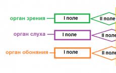
The analyzer is often called sensory system. Analyzers are classified according to the type of sensations in the formation of which they participate (see figure below).
Rice. Analyzers
This visual, auditory, vestibular, gustatory, olfactory, cutaneous, muscular and other analyzers. The analyzer has three sections:
- Peripheral department: a receptor designed to convert the energy of stimulation into the process of nervous excitation.
- Wiring department: a chain of centripetal (afferent) and intercalary neurons through which impulses are transmitted from receptors to the overlying parts of the central nervous system.
- Central department: a specific area of the cerebral cortex.
In addition to the ascending (afferent) pathways, there are descending fibers (efferent), through which the activity of the lower levels of the analyzer is regulated by its higher, especially cortical, sections.
| analyzer | peripheral section (sensory organ and receptors) | conductor department | central department |
|---|---|---|---|
| visual | retinal receptors | optic nerve | visual center in the occipital lobe of the KBP |
| auditory | sensory hair cells of the organ of Corti (spiral) organ of the cochlea | auditory nerve | auditory center in the temporal lobe |
| olfactory | olfactory receptors of the nasal epithelium | olfactory nerve | olfactory center in the temporal lobe |
| gustatory | taste buds of the oral cavity (mainly the root of the tongue) | glossopharyngeal nerve | taste center in the temporal lobe |
| tactile (tactile) | tactile corpuscles of the papillary dermis (pain, temperature, tactile and other receptors) | centripetal nerves; spinal cord, medulla oblongata, diencephalon | center of skin sensitivity in the central gyrus of the parietal lobe of the KBP |
| musculocutaneous | proprioceptors in muscles and ligaments | centripetal nerves; spinal cord; medulla oblongata and diencephalon | motor zone and adjacent areas of the frontal and parietal lobes. |
| vestibular | semicircular canaliculi and vestibule of the inner ear | vestibulocochlear nerve (VIII pair of cranial nerves) | cerebellum |
KBP*- cerebral cortex.
sense organs
A person has a number of important specialized peripheral formations - sense organs, providing the perception of external stimuli affecting the body.
The sense organ consists of receptors And auxiliary apparatus, which helps to capture, concentrate, focus, direct, etc. signal.
The sense organs include the organs of vision, hearing, smell, taste, and touch. By themselves they cannot provide sensation. For a subjective sensation to arise, it is necessary that the excitation that arises in the receptors enters the corresponding section of the cerebral cortex.
Structural fields of the cerebral cortex
If we consider the structural organization of the cerebral cortex, we can distinguish several fields with different cellular structures.
There are three main groups of fields in the cortex:
- primary
- secondary
- tertiary
Primary fields, or nuclear zones of the analyzers, are directly connected with the senses and organs of movement.
For example, the field of pain, temperature, musculocutaneous sensitivity in the posterior part of the central gyrus, the visual field in the occipital lobe, the auditory field in the temporal lobe and the motor field in the anterior part of the central gyrus.
Primary fields mature earlier than others in ontogenesis.
Function of primary fields: analysis of individual stimuli entering the cortex from the corresponding receptors.
When the primary fields are destroyed, so-called cortical blindness, cortical deafness, etc. occur.
Secondary fields located next to the primary ones and connected through them with the sense organs.
Function of secondary fields: generalization and further processing of incoming information. Individual sensations are synthesized in them into complexes that determine the processes of perception.
When secondary fields are damaged, a person sees and hears, but unable to comprehend understand the meaning of what you see and hear.
Both humans and animals have primary and secondary fields.
Tertiary fields, or overlap areas of analyzers, are located in the posterior half of the cortex - on the border of the parietal, temporal and occipital lobes and in the anterior parts of the frontal lobes. They occupy half of the entire area of the cerebral cortex and have numerous connections with all its parts.Most of the nerve fibers connecting the left and right hemispheres end in the tertiary fields.
Function of tertiary fields: organization of coordinated work of both hemispheres, analysis of all perceived signals, their comparison with previously received information, coordination of appropriate behavior,programming of motor activity.
These fields are found only in humans and mature later than other cortical fields.
The development of tertiary fields in humans is associated with the function of speech. Thinking (inner speech) is possible only with the joint activity of analyzers, the integration of information from which occurs in tertiary fields.
With congenital underdevelopment of the tertiary fields, a person is not able to master speech and even the simplest motor skills.

Rice. Structural fields of the cerebral cortex
Taking into account the location of the structural fields of the cerebral cortex, functional parts can be distinguished: sensory, motor and associative areas.
All sensory and motor areas occupy less than 20% of the surface of the cortex. The rest of the cortex constitutes the association region.
Association zones
Association zones- This functional areas cerebral cortex. They connect newly received sensory information with previously received and stored in memory blocks, and also compare information received from different receptors (see figure below).
Each associative area of the cortex is associated with several structural fields. The association zones include part of the parietal, frontal and temporal lobes. The boundaries of the associative zones are unclear; its neurons are involved in the integration of various information. Here comes the highest analysis and synthesis of irritations. As a result, complex elements of consciousness are formed.

Rice. Sulci and lobes of the cerebral cortex

Rice. Association areas of the cerebral cortex:
1. Ass motivating engine nal zone(frontal lobe)
2. Primary motor area
3. Primary somatosensory area
4. Parietal lobe of the cerebral hemispheres
5. Associative somatosensory (musculocutaneous) zone(parietal lobe)
6.Association visual area(occipital lobe)
7. Occipital lobe of the cerebral hemispheres
8. Primary visual area
9. Association auditory area(temporal lobes)
10. Primary auditory zone
11. Temporal lobe of the cerebral hemispheres
12. Olfactory cortex (inner surface of the temporal lobe)
13. Gustatory bark
14. Prefrontal association area
15. Frontal lobe of the cerebral hemispheres.
Sensory signals in the association zone are deciphered, interpreted and used to determine the most appropriate responses, which are transmitted to the associated motor (motor) zone.
Thus, associative zones are involved in the processes of memorization, learning and thinking, and the results of their activity constitute intelligence(the body’s ability to use acquired knowledge).
Individual large association areas are located in the cortex next to the corresponding sensory areas. For example, the visual association area is located in the occipital area immediately anterior to the sensory visual area and carries out complete processing of visual information.
Some association areas perform only part of the information processing and are connected to other association centers that perform further processing. For example, the auditory association area analyzes sounds, categorizing them, and then transmits signals to more specialized areas, such as the speech association area, where the meaning of words heard is perceived.
These zones belong to association cortex and participate in the organization of complex forms of behavior.
In the cerebral cortex, areas with less defined functions are distinguished. Thus, a significant part of the frontal lobes, especially on the right side, can be removed without noticeable damage. However, if a bilateral removal of the frontal areas is performed, severe mental disorders occur.
taste analyzer
Taste analyzer responsible for the perception and analysis of taste sensations.
Peripheral department: receptors - taste buds in the mucous membrane of the tongue, soft palate, tonsils and other organs of the oral cavity.


Rice. 1. Taste bud and taste bud
Taste buds bear taste buds on the lateral surface (Fig. 1, 2), which include 30 - 80 sensitive cells. Taste cells are dotted at their ends with microvilli - taste hairs. They come to the surface of the tongue through the taste pores. Taste cells continually divide and continually die. The replacement of cells located in the front part of the tongue, where they lie more superficially, occurs especially quickly.

Rice. 2. Taste bud: 1 - nerve taste fibers; 2 - taste bud (calyx); 3 - taste cells; 4 - supporting (supporting) cells; 5 - taste time

Rice. 3. Taste zones of the tongue: sweet - tip of the tongue; bitter - the base of the tongue; sour - lateral surface of the tongue; salty - tip of the tongue.
Taste sensations are caused only by substances dissolved in water.
Wiring department: fibers of the facial and glossopharyngeal nerve (Fig. 4).
Central department: inner side of the temporal lobe of the cerebral cortex.

olfactory analyzer
Olfactory analyzer responsible for the perception and analysis of smell.
- eating behavior;
- food testing for edibility;
- setting up the digestive system to process food (according to the mechanism of a conditioned reflex);
- defensive behavior (including manifestations of aggression).

Peripheral department: receptors in the mucous membrane of the upper part of the nasal cavity. Olfactory receptors in the nasal mucosa end in olfactory cilia. Gaseous substances dissolve in the mucus surrounding the cilia, then a nerve impulse arises as a result of a chemical reaction (Fig. 5).
Wiring department: olfactory nerve.
Central department: olfactory bulb (the structure of the forebrain in which information is processed) and the olfactory center located on the lower surface of the temporal and frontal lobes of the cerebral cortex (Fig. 6).
In the cortex, the smell is detected and the body’s adequate response to it is formed.

The perception of taste and smell complement each other, giving a holistic picture of the appearance and quality of food. Both analyzers are connected to the salivary center of the medulla oblongata and participate in the body’s nutritional reactions.
The tactile and muscle analyzers are combined into somatosensory system- system of musculoskeletal sensitivity.
Structure of the somatosensory analyzer
Peripheral department: proprioceptors of muscles and tendons; skin receptors ( mechanoreceptors, thermoreceptors, etc.).
Wiring department: afferent (sensitive) neurons; ascending tracts of the spinal cord; medulla oblongata, diencephalon nuclei.
Central department: sensory area in the parietal lobe of the cerebral cortex.
Skin receptors
The skin is the largest sensory organ in the human body. Many receptors are concentrated on its surface (about 2 m2).
Most scientists tend to believe that there are four main types of skin sensitivity: tactile, thermal, cold and pain.
Receptors are distributed unevenly and at different depths. Most of the receptors are in the skin of the fingers, palms, soles, lips and genitals.
MECHANORECEPTORS OF THE SKIN
- thin nerve fiber endings, entwining blood vessels, hair follicles, etc.
- Merkel cells- nerve endings of the basal layer of the epidermis (many on the fingertips);
- tactile Meissner corpuscles- complex receptors of the papillary dermis (many on the fingers, palms, soles, lips, tongue, genitals and nipples of the mammary glands);
- lamellar bodies- pressure and vibration receptors; located in the deep layers of the skin, in tendons, ligaments and mesentery;
- bulbs (Krause flasks)- nerve receptors inconnective tissue layer of mucous membranes, under the epidermis and among the muscle fibers of the tongue.
MECHANISM OF OPERATION OF MECHANORECEPTORS
Mechanical stimulus - deformation of the receptor membrane - decrease in the electrical resistance of the membrane - increase in membrane permeability for Na+ - depolarization of the receptor membrane - propagation of the nerve impulse
ADAPTATION OF SKIN MECHANORECEPTORS
- rapidly adapting receptors: skin mechanoreceptors in hair follicles, lamellar bodies (we do not feel the pressure of clothing, contact lenses, etc.);
- slow adapting receptors:tactile Meissner corpuscles.
The sensation of touch and pressure on the skin is quite accurately localized, that is, a person relates to a specific area of the skin surface. This localization is developed and consolidated in ontogenesis with the participation of vision and proprioception.
A person’s ability to separately perceive touch on two adjacent points of the skin also differs greatly in different areas of the skin. On the mucous membrane of the tongue, the threshold of spatial difference is 0.5 mm, and on the skin of the back - more than 60 mm.
Temperature reception
The human body temperature fluctuates within relatively narrow limits, so information about the ambient temperature, necessary for the functioning of thermoregulation mechanisms, is especially important.
Thermoreceptors are located in the skin, cornea, mucous membranes, and also in the central nervous system (hypothalamus).
TYPES OF THERMORECEPTORS
- cold thermoreceptors: numerous; lie close to the surface.
- thermal thermoreceptors: there are significantly fewer of them; lie in a deeper layer of skin.
- specific thermoreceptors: perceive only temperature;
- nonspecific thermoreceptors: perceive temperature and mechanical stimuli.
Thermoreceptors respond to temperature changes by increasing the frequency of generated impulses, which last steadily throughout the duration of the stimulus. A temperature change of 0.2 °C causes long-term changes in their impulses.
Under some conditions, cold receptors can be excited by heat, and thermal receptors by cold. This explains the acute sensation of cold when quickly immersed in a hot bath or the scalding effect of ice water.
The initial temperature sensations depend on the difference in skin temperature and the temperature of the active stimulus, its area and place of application. So, if the hand was held in water at a temperature of 27 °C, then at the first moment when the hand is transferred to water heated to 25 °C, it seems cold, but after a few seconds a true assessment of the absolute temperature of the water becomes possible.
Pain reception
Pain sensitivity is of paramount importance for the survival of the body, being a signal of danger under strong influences of various factors.
Impulses from pain receptors often indicate pathological processes in the body.
At the moment, no specific pain receptors have been found.
Two hypotheses about the organization of pain perception have been formulated:
- Exist specific pain receptors - free nerve endings with a high reaction threshold;
- Specific pain receptors does not exist; pain occurs when any receptors are overly stimulated.
The mechanism of receptor excitation during painful stimuli has not yet been clarified.
The most common cause of pain can be considered a change in H+ concentration due to toxic effects on respiratory enzymes or damage to cell membranes.
One of the possible causes of prolonged burning pain may be the release of histamine, proteolytic enzymes and other substances that cause a chain of biochemical reactions leading to excitation of nerve endings when cells are damaged.
Pain sensitivity is practically not represented at the cortical level, therefore the highest center of pain sensitivity is the thalamus, where 60% of neurons in the corresponding nuclei clearly react to painful stimulation.
ADAPTATION OF PAIN RECEPTORS
Adaptation of pain receptors depends on numerous factors and its mechanisms are poorly understood.
For example, a splinter, being motionless, does not cause much pain. Elderly people in some cases “get used to not noticing” headaches or joint pain.
However, in many cases, pain receptors do not show significant adaptation, which makes the patient’s suffering especially long and painful and requires the use of analgesics.
Painful stimuli cause a number of reflex somatic and autonomic reactions. When moderately expressed, these reactions have adaptive significance, but can lead to severe pathological effects, such as shock. Among these reactions are an increase in muscle tone, heart rate and respiration, an increase or decrease in blood pressure, constriction of the pupils, an increase in blood glucose and a number of other effects.
LOCALIZATION OF PAIN SENSITIVITY
In case of painful effects on the skin, a person localizes them quite accurately, but in case of diseases of the internal organs they may arise. referred pain. For example, with renal colic, patients complain of “incoming” sharp pain in the legs and rectum. There may also be reverse effects.
proprioception
Types of proprioceptors:
- neuromuscular spindles: provide information about the speed and force of muscle stretch and contraction;
- Golgi tendon receptors: provide information about the force of muscle contraction.
Functions of proprioceptors:
- perception of mechanical irritations;
- perception of the spatial arrangement of body parts.
NEUROMUSCULAR SPINDLE
Neuromuscular spindle- a complex receptor that includes modified muscle cells, afferent and efferent nerve processes and controls both the speed and degree of contraction and stretching of skeletal muscles.
The neuromuscular spindle is located deep within the muscle. Each spindle is covered with a capsule. Inside the capsule there is a bundle of special muscle fibers. The spindles are located parallel to the fibers of skeletal muscles, so when the muscle is stretched, the load on the spindles increases, and when it contracts, it decreases.

Rice. Neuromuscular spindle
GOLGI TENDON RECEPTORS
They are located in the area where the muscle fibers connect with the tendon.
Tendon receptors react weakly to muscle stretching, but are excited when it contracts. The intensity of their impulses is approximately proportional to the force of muscle contraction.

Rice. Golgi tendon receptor
JOINT RECEPTORS
They have been studied less than muscle ones. It is known that articular receptors respond to the position of the joint and to changes in the joint angle, thus participating in the feedback system from the motor system and in its control.
The visual analyzer includes:
- peripheral: retinal receptors;
- conduction section: optic nerve;
- central section: occipital lobe of the cerebral cortex.
Visual analyzer function: perception, conduction and decoding of visual signals.
Structures of the eye
The eye consists of eyeball And auxiliary apparatus.
Accessory eye apparatus
- brows- protection from sweat;
- eyelashes- protection from dust;
- eyelids- mechanical protection and moisture maintenance;
- lacrimal glands- located at the upper part of the outer edge of the orbit. It secretes tear fluid that moisturizes, washes and disinfects the eye. Excess tear fluid is removed into the nasal cavity through tear duct located in the inner corner of the orbit .
EYEBALL
The eyeball is roughly spherical in shape with a diameter of about 2.5 cm.
It is located on a cushion of fatin the anterior part of the orbit.
The eye has three membranes:
- tunica albuginea ( sclera) with a transparent cornea- outer very dense fibrous membrane of the eye;
- choroid with outer iris and ciliary body- penetrated by blood vessels (nutrition of the eye) and contains a pigment that prevents the scattering of light through the sclera;
- retina (retina) - inner lining of the eyeball -receptor part of the visual analyzer; function: direct perception of light and transmission of information to the central nervous system.

Conjunctiva- mucous membrane connecting the eyeball to the skin.
Tunica albuginea (sclera)- durable outer shell of the eye; the inner part of the sclera is impenetrable to set rays. Function: eye protection from external influences and light insulation;
Cornea- anterior transparent part of the sclera; is the first lens on the path of light rays. Function: mechanical protection of the eye and transmission of light rays.
Lens- a biconvex lens located behind the cornea. Function of the lens: focusing light rays. The lens has no blood vessels or nerves. Inflammatory processes do not develop in it. It contains many proteins, which can sometimes lose their transparency, leading to a disease called cataract.
Choroid- the middle layer of the eye, rich in blood vessels and pigment.
Iris- anterior pigmented part of the choroid; contains pigments melanin And lipofuscin, determining eye color.
Pupil- a round hole in the iris. Function: regulation of light flow entering the eye. Pupil diameter changes involuntarily with the help of smooth muscles of the iriswhen lighting changes.
Front and rear cameras- space in front and behind the iris filled with clear liquid ( aqueous humor).
Ciliary (ciliary) body- part of the middle (choroid) membrane of the eye; function: fixation of the lens, ensuring the process of accommodation (change in curvature) of the lens; production of aqueous humor in the chambers of the eye, thermoregulation.
Vitreous body- the cavity of the eye between the lens and the fundus of the eye , filled with a transparent viscous gel that maintains the shape of the eye.
Retina (retina)- receptor apparatus of the eye.
STRUCTURE OF THE RETINA
The retina is formed by the branches of the endings of the optic nerve, which, approaching the eyeball, passes through the tunica albuginea, and the sheath of the nerve merges with the tunica albuginea of the eye. Inside the eye, the nerve fibers are distributed in the form of a thin mesh membrane that lines the back 2/3 of the inner surface of the eyeball.
The retina is made up of supporting cells that form a mesh-like structure, hence its name. Only its back part perceives light rays. The retina, in its development and function, is part of the nervous system. However, the remaining parts of the eyeball play a supporting role in the retina’s perception of visual stimuli.
Retina- this is the part of the brain that is pushed outward, closer to the surface of the body, and maintains a connection with it through a pair of optic nerves.
Nerve cells form chains in the retina consisting of three neurons (see figure below):
- the first neurons have dendrites in the form of rods and cones; these neurons are the terminal cells of the optic nerve; they perceive visual stimuli and are light receptors.
- the second - bipolar neurons;
- the third are multipolar neurons ( ganglion cells); Axons extend from them, which stretch along the bottom of the eye and form the optic nerve.

Photosensitive elements of the retina:
- sticks- perceive brightness;
- cones- perceive color.
The cones are excited slowly and only by bright light. They are able to perceive color. There are three types of cones in the retina. The first perceive the color red, the second - green, the third - blue. Depending on the degree of excitation of the cones and the combination of irritations, the eye perceives different colors and shades.
The rods and cones in the retina of the eye are mixed together, but in some places they are very densely located, in others they are rare or absent altogether. For each nerve fiber there are approximately 8 cones and about 130 rods.
In area macular spot There are no rods on the retina - only cones; here the eye has the greatest visual acuity and the best color perception. Therefore, the eyeball is in continuous motion, so that the part of the object being examined falls on the macula. As you move away from the macula, the density of the rods increases, but then decreases.
In low light, only rods are involved in the vision process (twilight vision), and the eye does not distinguish colors, vision turns out to be achromatic (colorless).
Nerve fibers extend from the rods and cones, which unite to form the optic nerve. The place where the optic nerve exits the retina is called optic disc. There are no photosensitive elements in the area of the optic nerve head. Therefore, this place does not give a visual sensation and is called blind spot.
EYE MUSCLES
- oculomotor muscles- three pairs of striated skeletal muscles that are attached to the conjunctiva; carry out movement of the eyeball;
- pupil muscles- smooth muscles of the iris (circular and radial), changing the diameter of the pupil;
The circular muscle (contractor) of the pupil is innervated by parasympathetic fibers from the oculomotor nerve, and the radial muscle (dilator) of the pupil is innervated by fibers of the sympathetic nerve. The iris thus regulates the amount of light entering the eye; in strong, bright light, the pupil narrows and limits the entry of rays, and in weak light, it expands, allowing more rays to penetrate. The diameter of the pupil is influenced by the hormone adrenaline. When a person is in an excited state (fear, anger, etc.), the amount of adrenaline in the blood increases, and this causes the pupil to dilate.
The movements of the muscles of both pupils are controlled from one center and occur synchronously. Therefore, both pupils always dilate or contract equally. Even if you apply bright light to only one eye, the pupil of the other eye also narrows. - lens muscles(ciliary muscles) - smooth muscles that change the curvature of the lens ( accommodation--focusing the image on the retina).
Wiring department
The optic nerve conducts light stimuli from the eye to the visual center and contains sensory fibers.
Moving away from the posterior pole of the eyeball, the optic nerve leaves the orbit and, entering the cranial cavity, through the optic canal, together with the same nerve on the other side, forms a chiasm ( chiasmus) under the hypolalamus. After the chiasm, the optic nerves continue in visual tracts. The optic nerve is connected to the nuclei of the diencephalon, and through them to the cerebral cortex.
Each optic nerve contains the totality of all the processes of the nerve cells of the retina of one eye. In the area of the chiasm, an incomplete crossover of fibers occurs, and each optic tract contains about 50% of the fibers of the opposite side and the same number of fibers of the same side.

Central department
The central section of the visual analyzer is located in the occipital lobe of the cerebral cortex.
Impulses from light stimuli travel along the optic nerve to the cerebral cortex of the occipital lobe, where the visual center is located.
The fibers of each nerve are connected to the two hemispheres of the brain, and the image obtained on the left half of the retina of each eye is analyzed in the visual cortex of the left hemisphere, and on the right half of the retina - in the cortex of the right hemisphere.

visual impairment
With age and under the influence of other reasons, the ability to control the curvature of the lens surface weakens.
Myopia (myopia)- focusing the image in front of the retina; develops due to an increase in the curvature of the lens, which can occur due to improper metabolism or poor visual hygiene. AND use glasses with concave lenses.
Farsightedness- focusing the image behind the retina; occurs due to a decrease in the convexity of the lens. ANDcope with glasseswith convex lenses.

There are two ways to conduct sounds:
- air conduction: through the external auditory canal, eardrum and chain of auditory ossicles;
- tissue conductivity b: through the tissues of the skull.
Function of the auditory analyzer: perception and analysis of sound stimuli.
Peripheral: auditory receptors in the inner ear cavity.
Conductor section: auditory nerve.
Central division: auditory zone in the temporal lobe of the cerebral cortex.


Rice. Temporal bone Fig. Location of the hearing organ in the cavity of the temporal bone
ear structure
The human hearing organ is located in the cranial cavity in the thickness of the temporal bone.
It is divided into three sections: the outer, middle and inner ear. These departments are closely connected anatomically and functionally.
Outer ear consists of the external auditory canal and the auricle.
Middle ear- tympanic cavity; it is separated from the outer ear by the eardrum.
Inner ear, or labyrinth, - the section of the ear where irritation of the receptors of the auditory (cochlear) nerve occurs; it is placed inside the pyramid of the temporal bone. The inner ear forms the organ of hearing and balance.
The outer and middle ears are of secondary importance: they conduct sound vibrations to the inner ear, and are thus a sound-conducting apparatus.

Rice. Ear sections
EXTERNAL EAR
Outer ear includes auricle And external auditory canal, which are designed to capture and conduct sound vibrations.
Auricle formed by three tissues:
- a thin plate of hyaline cartilage, covered on both sides with perichondrium, having a complex convex-concave shape that determines the relief of the auricle;
- the skin is very thin, tightly adjacent to the perichondrium and has almost no fatty tissue;
- subcutaneous fatty tissue, located in significant quantities in the lower part of the auricle - earlobe.
The auricle is attached to the temporal bone by ligaments and has vestigial muscles that are well defined in animals.
The auricle is designed to concentrate sound vibrations as much as possible and direct them into the external auditory opening.
The shape, size, position of the auricle and the size of the ear lobe are individual for each person.
Darwin's tubercle- a rudimentary triangular protrusion, which is observed in 10% of people in the superior-posterior region of the conchal helix; it corresponds to the top of the animal's ear.

Rice. Darwin's tubercle
External auditory passage is an S-shaped tube approximately 3 cm long and 0.7 cm in diameter, which opens externally with the auditory opening and is separated from the middle ear cavity eardrum.
The cartilaginous part, which is a continuation of the cartilage of the auricle, makes up 1/3 of its length, the remaining 2/3 is formed by the bone canal of the temporal bone. At the point where the cartilaginous section transitions into the bone canal, it narrows and bends. In this place there is a ligament of elastic connective tissue. This structure makes it possible to stretch the cartilaginous part of the passage in length and width.
In the cartilaginous part of the ear canal, the skin is covered with short hairs that protect against small particles from entering the ear. The sebaceous glands open into the hair follicles. Characteristic of the skin of this section is the presence of sulfur glands in the deeper layers.
Sulfur glands are derivatives of the sweat glands. Sulfur glands drain either into the hair follicles or freely into the skin. The sulfur glands secrete a light yellow secretion, which, together with the secretion of the sebaceous glands and the rejected epithelium, forms earwax.
Earwax- light yellow secretion of the sulfur glands of the external auditory canal.
Sulfur consists of proteins, fats, fatty acids and mineral salts. Some proteins are immunoglobulins that determine the protective function. In addition, sulfur contains dead cells, sebum, dust and other inclusions.
Function of earwax:
- moisturizing the skin of the external auditory canal;
- cleaning the ear canal from foreign particles (dust, litter, insects);
- protection against bacteria, fungi and viruses;
- grease in the outer part of the ear canal prevents water from entering it.
Earwax, along with impurities, is naturally removed from the ear canal through chewing movements and speech. In addition, the skin of the ear canal is constantly renewed and grows outward from the ear canal, taking wax with it.
Interior bone section The external auditory canal is a canal of the temporal bone that ends in the eardrum. In the middle of the bone section there is a narrowing of the auditory canal - the isthmus, behind which there is a wider area.
The skin of the bony part is thin, does not contain hair follicles and glands and extends onto the eardrum, forming its outer layer.
Eardrum represents thin oval (11 x 9 mm) translucent plate, impermeable to water and air. Membraneconsists of elastic and collagen fibers, which in its upper part are replaced by fibers of loose connective tissue.On the side of the auditory canal, the membrane is covered with squamous epithelium, and on the side of the tympanic cavity - with mucosal epithelium.
In the central part, the eardrum is concave; the handle of the malleus, the first auditory ossicle of the middle ear, is attached to it from the side of the tympanic cavity.
The eardrum begins and develops along with the organs of the outer ear.
MIDDLE EAR
The middle ear includes a mucous membrane lined and filled with air tympanic cavity(volume about 1 Withm3 cm3), three auditory ossicles and auditory (Eustachian) tube.

Rice. Middle ear
Tympanic cavity located in the thickness of the temporal bone, between the eardrum and the bony labyrinth. The tympanic cavity contains the auditory ossicles, muscles, ligaments, blood vessels and nerves. The walls of the cavity and all the organs located in it are covered with a mucous membrane.
In the septum separating the tympanic cavity from the inner ear, there are two windows:
- oval window: located in the upper part of the septum, leads to the vestibule of the inner ear; closed by the base of the stapes;
- round window: located in lower part of the septum, leads to the beginning of the cochlea; closed by the secondary tympanic membrane.
There are three auditory ossicles in the tympanic cavity: malleus, incus and stapes (= stapes). The auditory ossicles are small. Connecting with each other, they form a chain that stretches from the eardrum to the oval opening. All bones are connected to each other using joints and are covered with a mucous membrane.
Hammer the handle is fused with the eardrum, and the head is connected to the anvil, which in turn is movably connected to stirrup. The base of the stapes covers the oval window of the vestibule.
The muscles of the tympanic cavity (tensor tympani and stapedius) keep the auditory ossicles in a state of tension and protect the inner ear from excessive sound stimulation.
Auditory (Eustachian) tube connects the tympanic cavity of the middle ear with the nasopharynx. This a muscular tube that opens when swallowing and yawning.
The mucous membrane lining the auditory tube is a continuation of the mucous membrane of the nasopharynx and consists of ciliated epithelium with the movement of cilia from the tympanic cavity into the nasopharynx.
Functions of the Eustachian tube:
- balancing the pressure between the tympanic cavity and the external environment to maintain normal operation of the sound-conducting apparatus;
- protection against infections;
- removal of accidentally penetrated particles from the tympanic cavity.
INNER EAR
The inner ear consists of a bony labyrinth and a membranous labyrinth inserted into it.
Bone labyrinth consists of three departments: vestibule, cochlea And three semicircular canals.
vestibule- a cavity of small size and irregular shape, on the outer wall of which there are two windows (round and oval) leading into the tympanic cavity. The anterior part of the vestibule communicates with the cochlea through the scala vestibule. The back part contains two impressions for the vestibular sacs.
Snail- bone spiral channel of 2.5 turns. The axis of the cochlea lies horizontally and is called the bony cochlear shaft. A bone spiral plate wraps around the rod, which partially blocks the spiral canal of the cochlea and divides it on staircase vestibule And staircase drum. They communicate with each other only through a hole located at the top of the cochlea.
Rice. Structure of the cochlea: 1 - basement membrane; 2 - organ of Corti; 3 - Reisner membrane; 4 - staircase vestibule; 5 - spiral ganglion; 6 - scala tympani; 7 - vestibular-helical nerve; 8 - spindle.
Semicircular canals- bone formations located in three mutually perpendicular planes. Each channel has an expanded stalk (ampule).

Rice. Cochlea and semicircular canals
Membranous labyrinth filled endolymph And consists of three departments:
- membranous snail, orcochlear duct,continuation of the spiral plate between the scala vestibule and the scala tympani. The cochlear duct contains auditory receptors -spiral, or organ of Corti;
- three semicircular canals and two pouches located in the vestibule, which play the role of the vestibular apparatus.
Between the bony and membranous labyrinth there is perilymph--modified cerebrospinal fluid.
organ of corti
On the plate of the cochlear duct, which is a continuation of the bony spiral plate, there is organ of Corti (spiral).
The spiral organ is responsible for the perception of sound stimuli. It acts as a microphone, transforming mechanical vibrations into electrical ones.
The organ of Corti consists of supporting and sensory hair cells.
Rice. Organ of Corti
Hair cells have hairs that rise above the surface and reach the integumentary membrane (tectorial membrane). The latter extends from the edge of the spiral bone plate and hangs over the organ of Corti.
When sound stimulation of the inner ear occurs, vibrations occur in the main membrane on which the hair cells are located. Such vibrations cause stretching and compression of the hairs against the integumentary membrane, and generate a nerve impulse in the sensory neurons of the spiral ganglion.

Rice. Hair cells
WIRING DEPARTMENT
The nerve impulse from the hair cells spreads to the spiral ganglion.
Then by auditory ( vestibulocochlear) nerve the impulse enters the medulla oblongata.
In the pons, some of the nerve fibers pass through the decussation (chiasm) to the opposite side and go to the quadrigeminal region of the midbrain.
Nerve impulses through the nuclei of the diencephalon are transmitted to the auditory zone of the temporal lobe of the cerebral cortex.
The primary auditory centers serve for the perception of auditory sensations, the secondary ones for their processing (understanding speech and sounds, perceiving music).
Rice. Hearing analyzer
The facial nerve passes along with the auditory nerve into the inner ear and under the mucous membrane of the middle ear follows to the base of the skull. It can be easily damaged by inflammation of the middle ear or trauma to the skull, so hearing and balance disorders are often accompanied by paralysis of the facial muscles.
Physiology of hearing
The hearing function of the ear is provided by two mechanisms:
- sound conduction: conduction of sounds through the outer and middle ear to the inner ear;
- sound perception: perception of sounds by receptors of the organ of Corti.
SOUND CONDUCTION
The outer and middle ear and the perilymph of the inner ear belong to the sound-conducting apparatus, and the inner ear, that is, the spiral organ and leading nerve pathways, belong to the sound-receiving apparatus. The auricle, due to its shape, concentrates sound energy and directs it towards the external auditory canal, which conducts sound vibrations to the eardrum.
Having reached the eardrum, sound waves cause it to vibrate. These vibrations of the eardrum are transmitted to the malleus, through the joint to the incus, through the joint to the stapes, which closes the window of the vestibule (oval window). Depending on the phase of the sound vibrations, the base of the stapes is either squeezed into the labyrinth or pulled out of it. These movements of the stapes cause vibrations in the perilymph (see figure), which are transmitted to the main membrane of the cochlea and to the organ of Corti located on it.
As a result of vibrations of the main membrane, the hair cells of the spiral organ touch the integumentary (tentorial) membrane overhanging them. In this case, stretching or contraction of the hairs occurs, which is the main mechanism for converting the energy of mechanical vibrations into the physiological process of nervous excitation.
The nerve impulse is transmitted by the endings of the auditory nerve to the nuclei of the medulla oblongata. From here, impulses travel along corresponding leading paths to the auditory centers in the temporal parts of the cerebral cortex. Here the nervous excitement turns into a sensation of sound.
Rice. Sound path: auricle - external auditory canal - tympanic membrane - malleus - incus - pedicle - oval window - vestibule of the inner ear - scala vestibule - basement membrane - hair cells of the organ of Corti. Path of the nerve impulse: hair cells of the organ of Corti - spiral ganglion - auditory nerve - medulla oblongata - diencephalon nuclei - temporal lobe of the cerebral cortex.
SOUND PERCEPTION
A person perceives sounds of the external environment with an oscillation frequency from 16 to 20,000 Hz (1 Hz = 1 oscillation per 1 s).
High-frequency sounds are perceived by the lower part of the helix, and low-frequency sounds by its apex.
Rice. Schematic representation of the main membrane of the cochlea (frequencies distinguishable by different parts of the membrane are indicated)
Ototopics- WithThe ability to locate a sound source in cases where we cannot see it is called. It is associated with the symmetrical function of both ears and is regulated by the activity of the central nervous system. This ability arises because the sound that comes from the side does not enter different ears at the same time: into the ear of the opposite side - with a delay of 0.0006 s, with a different intensity and in a different phase. These differences in the perception of sound by different ears make it possible to determine the direction of the sound source.